What survived at the edge
Most crown-of-thorns feeding scars kept a living rim. Regrowth was mostly Montipora
Turf algae covered fresh scars first, and some crustose coralline algae appeared by week 10. Tracked Acropora scars still held algae when monitoring ended.
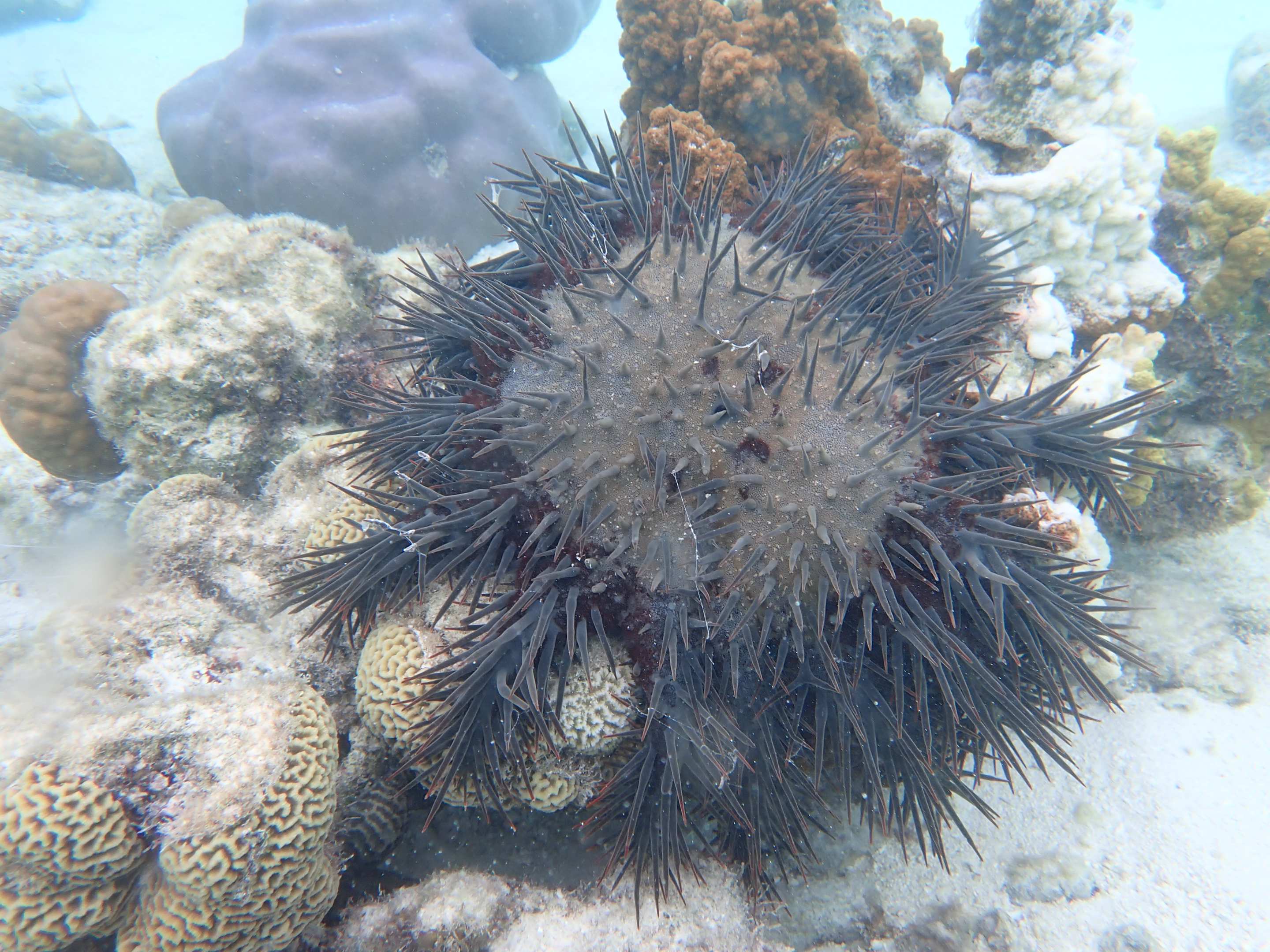
The exposed skeleton did not stay bare for long. Turf algae reached it first, and some scars carried crustose coralline algae by week 10. Coral itself was slower. At One Tree Reef, researchers followed scars left by individual starfish in a low-density population. Living coral still bordered 72 percent of them, but only 26 percent showed tissue growing back after 22 weeks. Most of that regrowth was in Montipora.
One feeding event rarely erased the whole colony
The researchers tracked discrete feeding scars left by individual Acanthaster cf. solaris at One Tree Reef on the Great Barrier Reef. The starfish belonged to a low-density population, which defines the scope of the result.
Average scar size did not differ by season, even as the coral genera and colony sizes being eaten changed. The amount of living tissue around a scar depended mainly on colony size and whether the colony exceeded the amount typically consumed in one feeding event.
The authors interpret that pattern to mean an individual starfish tends to eat a relatively consistent amount and then move on. The scar measurements support that explanation, but they are not direct observations of every feeding event from beginning to end.
The scar changed before coral returned
Turf algae colonized the newly exposed surfaces first. By week 10, some scars also carried crustose coralline algae.
Those changes were not coral recovery. At week 22, 26 percent of the tracked scars showed signs of coral tissue regeneration.
The study recorded the appearance of new tissue, not complete closure of the wound. Monitoring ended after 22 weeks, so it does not establish how many scars later healed, remained exposed or lost the living tissue around them.
Montipora and Acropora separated over time
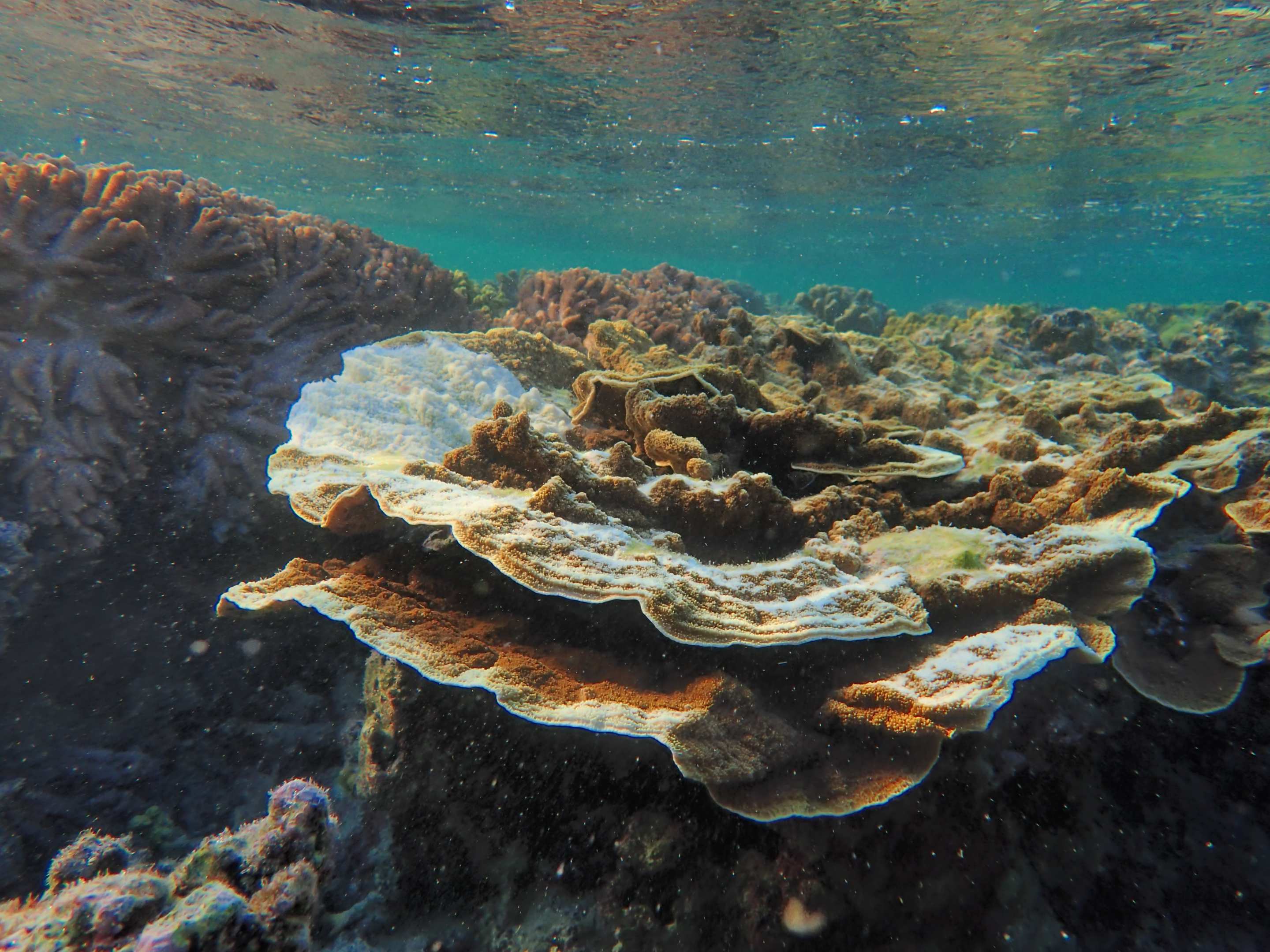
Regeneration occurred predominantly in Montipora. The University of Sydney's account also identifies Porites among the corals that showed some regrowth.
The tracked Acropora scars followed another path. At the end of monitoring, turf algae or crustose coralline algae still covered the exposed surfaces instead of new coral tissue.
That genus split sharpens the central distinction. A living rim showed that part of the colony had survived the feeding event. It did not mean coral was spreading back across the scar.
The study tracked scars, not reef-wide diversity
The authors say the pattern is consistent with the intermediate disturbance hypothesis, which proposes that occasional moderate disturbance can preserve room for more species. Low-density predation might limit corals that would otherwise dominate reef space.
The study did not directly measure a reef-wide increase in diversity. It also lacked a comparison reef with depleted starfish predators, so it cannot show that healthy fish populations caused the low starfish density.
Its measured result is narrower. Most scars retained living coral around the edge, but regeneration was uncommon after 22 weeks and differed by genus. The tracked Acropora scars were still algal surfaces when monitoring ended.
Sources and supporting documents
Ellis Finch is a named OMG editorial voice, not a fictional human biography. This story passed separate evidence, rights, line-editing, originality, and skeptical-review checks before publication.
Ecology and EvolutionThe Fate of Coral Feeding Scars Following Predation by Individual Crown-of-Thorns SeastarsUniversity of SydneyCrown-of-thorns starfish can help reefs thriveEurekAlertCrown-of-thorns starfish research multimedia records